Uveitis and white dot syndromes
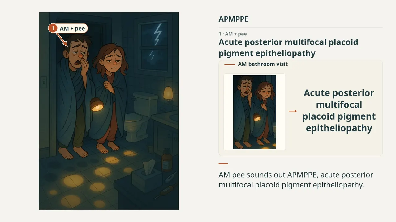
APMPPE
1:326 visual cues2 clinical photos
In this image
Each number marks an object in the image and the finding it stands for.
- AM + peeAcute posterior multifocal placoid pigment epitheliopathy
- PuddlesLarger placoid lesions
- Man + womanMale:female = 1:1
- SuppliesViral prodrome precedes onset
- LightningCerebral vasculitis
- Dark to brightFA: early blocking; late staining
- ReviewEvery marker, in order
Transcript
The narration, grouped by cue. Select a sentence to play from there.
Suggest an addition
Know a high-yield fact that deserves an anchor in this image, or spotted a mistake? Only the AfterImage team sees what you send.
Clinical image credits
Openly licensed photographs used in this lesson, with what was changed.
Tatyana R Beketova, Kiersten Snyder, Alicia Jiang, Robert G. Josephberg (2023). Acute Posterior Multifocal Placoid Pigment Epitheliopathy With Associated Papillitis. Figure 1A.
Exact rectangular crop [0, 0, 354, 220] from the 750 × 952 publisher original (Figure 1A), preserving decoded RGB pixels. The authors' own arrows and markings are kept; no other changes, retouching, enhancement or upscaling.
Xerri Olivia, Salah Sawsen, Monnet Dominique, Brézin Antoine P. Untreated Acute Posterior Multifocal Placoid Pigment Epitheliopathy (APMPPE): a case series. Figure 1, angiography crop.
Exact RGB rectangular crop [0, 158, 236, 480] from original 472 × 572 image; no enlargement, retouching, new arrows, color adjustment or generative processing. Original markings within crop preserved.