Retina and optic nerve
Stargardt disease
3:2312 visual cues3 clinical photosFree lesson
In this image
Each number marks an object in the image and the finding it stands for.
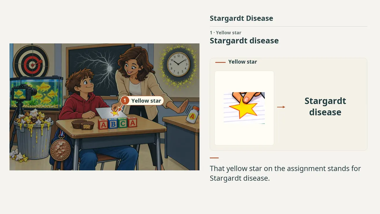
- Yellow starStargardt disease
- StudentJuvenile macular dystrophy
- DartboardBull’s-eye maculopathy
- GoldfishYellow pisciform flecks
- Overflowing trashLipofuscin accumulation
- Peanut butter cupUsually autosomal recessive
- BlocksABCA4
- BlackboardDark choroid on angiography
- CameraFundus autofluorescence
- Clean clock ringPeripapillary sparing
- Bronze medalBeaten-bronze appearance
- Vitamin AVitamin A supplementation
- ReviewEvery marker, in order
Transcript
The narration, grouped by cue. Select a sentence to play from there.
Suggest an addition
Know a high-yield fact that deserves an anchor in this image, or spotted a mistake? Only the AfterImage team sees what you send.
Clinical image credits
Openly licensed photographs used in this lesson, with what was changed.
Saket Y. Maheshwari; Swarupa Chakole. A Rare Occurrence of Stargardt Disease in a Quadragenarian Adult. Cureus 2022. Figure 3a.
Rectangular panel extraction only; no image retouching, recoloring, sharpening, generative processing, or clinical markers added. Original figure annotation and panel letters retained.
Saket Y. Maheshwari; Swarupa Chakole. A Rare Occurrence of Stargardt Disease in a Quadragenarian Adult. Cureus 2022. Figure 3b.
Rectangular panel extraction only; no image retouching, recoloring, sharpening, generative processing, or clinical markers added. Original figure annotation and panel letters retained.
