Retina and optic nerve
Central areolar choroidal dystrophy
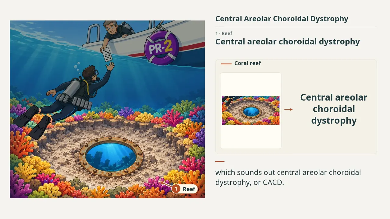
1:569 visual cues1 clinical photo
In this image
Each number marks an object in the image and the finding it stands for.
- ReefCentral areolar choroidal dystrophy
- Central zoneWell-defined central macular atrophy
- Surface coralProgressive RPE and outer retinal atrophy
- Fine networkChoriocapillaris atrophy; deeper structures visible
- PortholeFA window defect from RPE pigment loss
- DiverTypically symptomatic in adulthood
- 3 / 5 weightsCommonly symptomatic in the 3rd–5th decades
- PR-2PRPH2: classic genetic association
- DominoAutosomal dominant inheritance
- ReviewEvery marker, in order
Transcript
The narration, grouped by cue. Select a sentence to play from there.
Suggest an addition
Know a high-yield fact that deserves an anchor in this image, or spotted a mistake? Only the AfterImage team sees what you send.
Clinical image credits
Openly licensed photographs used in this lesson, with what was changed.
Eveline Kersten, Maartje J. Geerlings, Marc Pauper, Jordi Corominas, Bjorn Bakker, Lebriz Altay, Sascha Fauser, Eiko K. de Jong, Carel B. Hoyng, Anneke I. den Hollander (2018). Genetic screening for macular dystrophies in patients clinically diagnosed with dry age‐related macular degeneration. Figure 1E.
Exact rectangular crop [0, 568, 351, 820] from the 709 × 1079 publisher original (Figure 1E), preserving decoded RGB pixels. The authors' own arrows and markings are kept; no other changes, retouching, enhancement or upscaling.