Retina and optic nerve
Goldmann-Favre syndrome
3:4714 visual cues2 clinical photos
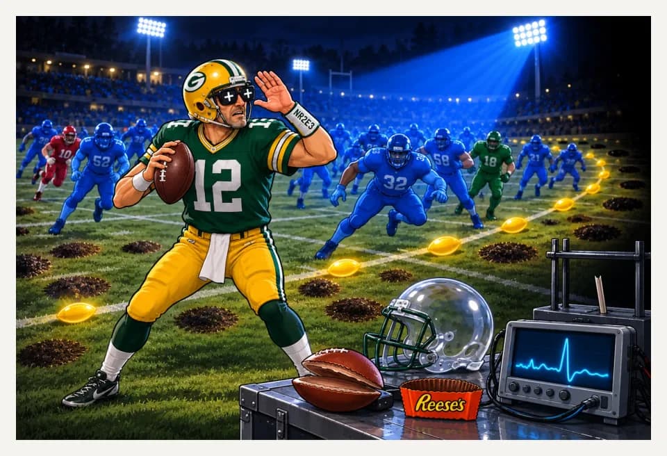
In this image
Each number marks an object in the image and the finding it stands for.
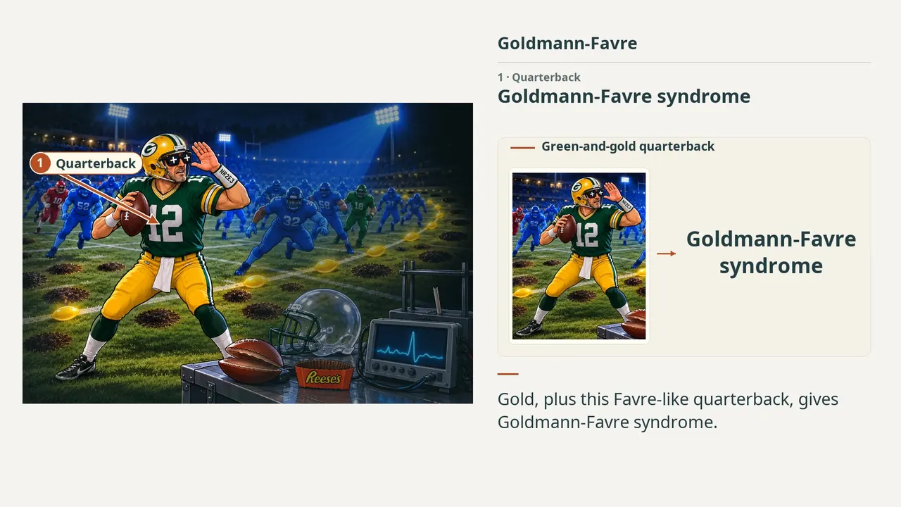
- QuarterbackGoldmann-Favre syndrome
- Night gameNyctalopia
- PlayersEnhanced S-cone syndrome · fewer red and green cones
- Rod rackFew, if any, functional rods
- Blue lightIncreased sensitivity to blue light
- Dark patchesPigmentary retinal degeneration
- Pigment discsNummular pigmentary deposits
- Yellow ballsYellow sheenlike lesions along the arcades
- Empty helmetOptically empty vitreous
- Split footballRetinoschisis
- Plus glassesHyperopia
- Dark sidelinesPeripheral to midperipheral field loss
- Reese's + NR2E3Autosomal recessive · NR2E3 mutation
- ERG testerPathognomonic ERG abnormalities
- ReviewEvery marker, in order
Transcript
The narration, grouped by cue. Select a sentence to play from there.
Suggest an addition
Know a high-yield fact that deserves an anchor in this image, or spotted a mistake? Only the AfterImage team sees what you send.
Clinical image credits
Openly licensed photographs used in this lesson, with what was changed.
Klaudia Szala and Bogumiła Wójcik-Niklewska (2025). A Rare Vitreoretinal Degenerative Disorder: Goldmann–Favre Syndrome Complicated with Choroidal Neovascularization in a Pediatric Patient.
Exact rectangular crop [0, 0, 374, 379] from 749 × 379 publisher figure. Decoded RGB pixels preserved; no enhancement, arrows, retouching or upscaling.
Klaudia Szala and Bogumiła Wójcik-Niklewska (2025). A Rare Vitreoretinal Degenerative Disorder: Goldmann–Favre Syndrome Complicated with Choroidal Neovascularization in a Pediatric Patient.
Exact rectangular crop [0, 0, 391, 267] from 785 × 532 publisher figure. Decoded RGB pixels preserved; no enhancement, arrows, retouching or upscaling.