Retina and optic nerve
Gyrate atrophy
2:0311 visual cues1 clinical photo
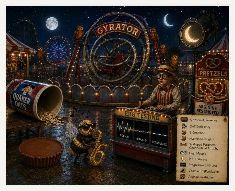
In this image
Each number marks an object in the image and the finding it stands for.
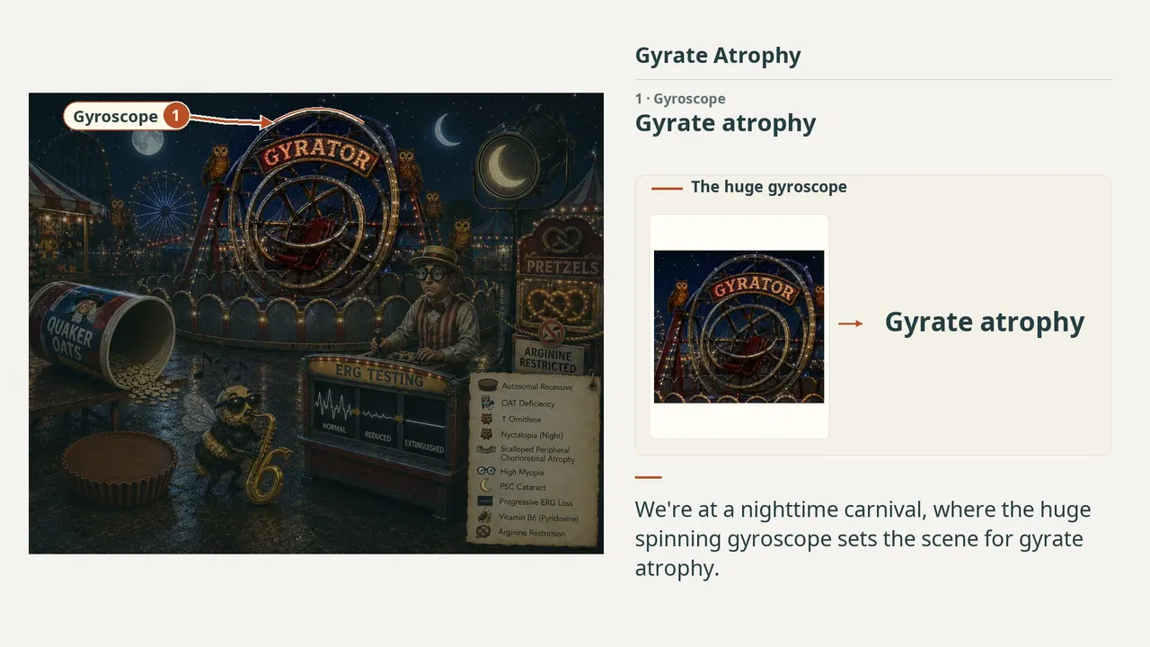
- GyroscopeGyrate atrophy
- Candy cupAutosomal recessive inheritance
- OatsOAT deficiency
- OwlsMarkedly elevated plasma ornithine
- FenceScalloped chorioretinal atrophy
- NightNyctalopia
- GlassesHigh myopia
- CrescentPSC cataract
- ERGProgressive ERG reduction
- Bee + 6Vitamin B6 supplementation
- PretzelsDietary arginine restriction
- ReviewEvery marker, in order
Transcript
The narration, grouped by cue. Select a sentence to play from there.
Suggest an addition
Know a high-yield fact that deserves an anchor in this image, or spotted a mistake? Only the AfterImage team sees what you send.
Clinical image credits
Openly licensed photographs used in this lesson, with what was changed.
Palmer E, Stepien KM, Campbell C, Barton S, Iosifidis C, Ghosh A, Broomfield A, Woodall A, Wilcox G, Sergouniotis PI, Black GC (2023). Clinical, biochemical and molecular analysis in a cohort of individuals with gyrate atrophy. Orphanet Journal of Rare Diseases 18:265. Figure 1A, left colour fundus photograph.
Cropped to the left colour panel; no other visual changes.
Exact changesHide exact changes
Exact rectangular crop [0, 0, 898, 545] from the 1770 × 1730 publisher JPEG. Retains the complete left colour fundus photograph from Figure 1A, its original A label and narrow original white margins. Excludes the right colour photograph and all Figure 1B and 1C panels. Losslessly encoded as RGB PNG with decoded RGB pixels identical to the corresponding original JPEG crop. No retouching, recoloring, enhancement, added annotations, arrows or upscaling.