Retina and optic nerve
Leber congenital amaurosis
LCA
1:579 visual cues2 clinical photos
In this image
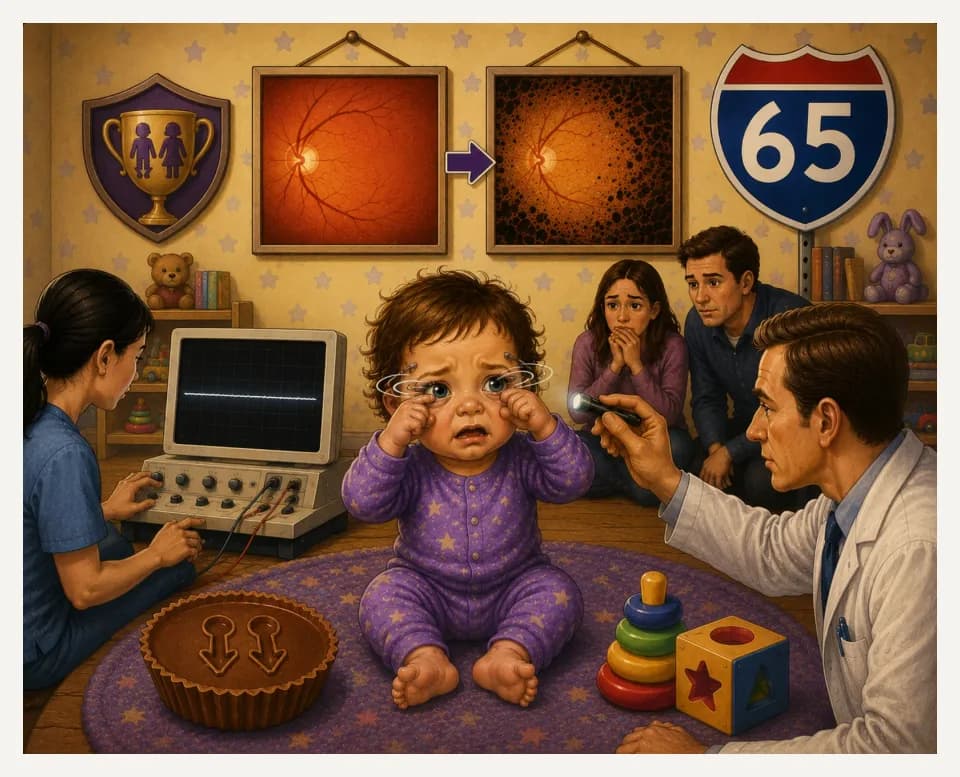
Each number marks an object in the image and the finding it stands for.
- BabyPresents in infancy
- Reeses cupAutosomal recessive inheritance
- Route 65RPE65 mutation
- Unused toysSevere vision loss at birth
- Eye rubbingOculodigital sign
- ShakingNystagmus
- PenlightNo constriction to light; Paradoxical pupillary response
- Fundus picturesInitially normal fundus; Progresses to an RP-like fundus
- Flat lineTotally extinguished ERG
- ReviewEvery marker, in order
Transcript
The narration, grouped by cue. Select a sentence to play from there.
Suggest an addition
Know a high-yield fact that deserves an anchor in this image, or spotted a mistake? Only the AfterImage team sees what you send.
Clinical image credits
Openly licensed photographs used in this lesson, with what was changed.
Neruban Kumaran, Anthony T. Moore, Richard G. Weleber, Michel Michaelides (2017). Leber congenital amaurosis/early-onset severe retinal dystrophy: clinical features, molecular genetics and therapeutic interventions.
Exact rectangular crop [1191, 0, 1772, 492] from 1772 × 492 publisher figure. Decoded RGB pixels preserved; no enhancement, arrows, retouching or upscaling.
Natsuki Higa, Takaaki Hayashi, Kei Mizobuchi, Maki Iwasa, Shingo Kubota, Kazuki Kuniyoshi, Shuhei Kameya, Hiroyuki Kondo, Mineo Kondo, Tadashi Nakano (2024). A novel RPE65 variant p.(Ala391Asp) in Leber congenital amaurosis: a case report and literature review in Japan.
Exact rectangular crop [0, 0, 1535, 1304] from 1535 × 1304 publisher figure. Decoded RGB pixels preserved; no enhancement, arrows, retouching or upscaling.