Uveitis and white dot syndromes
Punctate inner choroidopathy
PIC
1:305 visual cues2 clinical photos
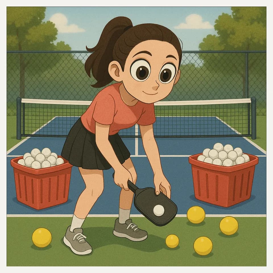
In this image
Each number marks an object in the image and the finding it stands for.
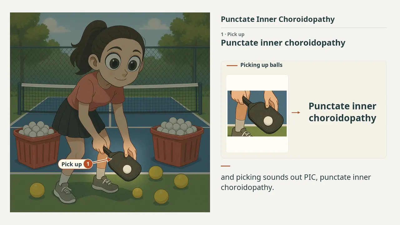
- Pick upPunctate inner choroidopathy
- EyesYoung myopic women
- Collected ballsNo vitritis
- Yellow ballsRPE disruption to chorioretinal scars
- Red binsCNVM: 50% of eyes; more common at presentation than MCP
- ReviewEvery marker, in order
Transcript
The narration, grouped by cue. Select a sentence to play from there.
Suggest an addition
Know a high-yield fact that deserves an anchor in this image, or spotted a mistake? Only the AfterImage team sees what you send.
Clinical image credits
Openly licensed photographs used in this lesson, with what was changed.
Evianne L. de Groot, Jeannette Ossewaarde–van Norel, Joke H. de Boer, Sanne Hiddingh, Bjorn Bakker, Ramon A. C. van Huet, Ninette H. ten Dam–van Loon, Alberta A. H. J. Thiadens, Magda A. Meester-Smoor, Yvonne de Jong–Hesse, Leonoor I. Los, Anneke I. den Hollander, Camiel J. F. Boon, Lambertus A. Kiemeney, Kristel R. van Eijk, Mark K. Bakker, Carel B. Hoyng, Jonas J. W. Kuiper (2023). Association of Risk Variants in the CFH Gene With Elevated Levels of Coagulation and Complement Factors in Idiopathic Multifocal Choroiditis. Figure 1B.
Exact rectangular crop [394, 49, 781, 355] from the 783 × 357 publisher original (Figure 1B), preserving decoded RGB pixels. The authors' own arrows and markings are kept; no other changes, retouching, enhancement or upscaling.
Aniruddha Agarwal, Sabia Handa, Alessandro Marchese, Salvatore Parrulli, Alessandro Invernizzi, Roel J. Erckens, Tos T. J. M. Berendschot, C. A. B. Webers, Reema Bansal, Vishali Gupta. Optical Coherence Tomography Findings of Underlying Choroidal Neovascularization in Punctate Inner Choroidopathy. Figure 2, cnv crop.
Exact RGB rectangular crop [26, 428, 330, 714] from original 1772 × 1511 image; no enlargement, retouching, new arrows, color adjustment or generative processing. Original markings within crop preserved.