Uveitis and white dot syndromes
Serpiginous choroiditis
1:398 visual cues1 clinical photo
In this image
Each number marks an object in the image and the finding it stands for.
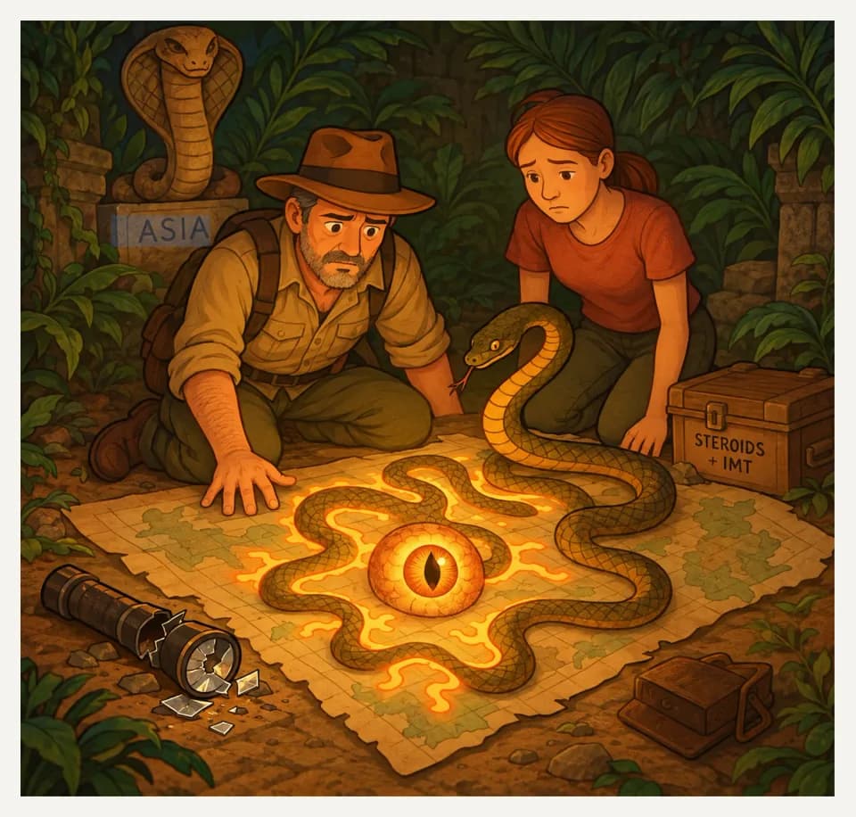
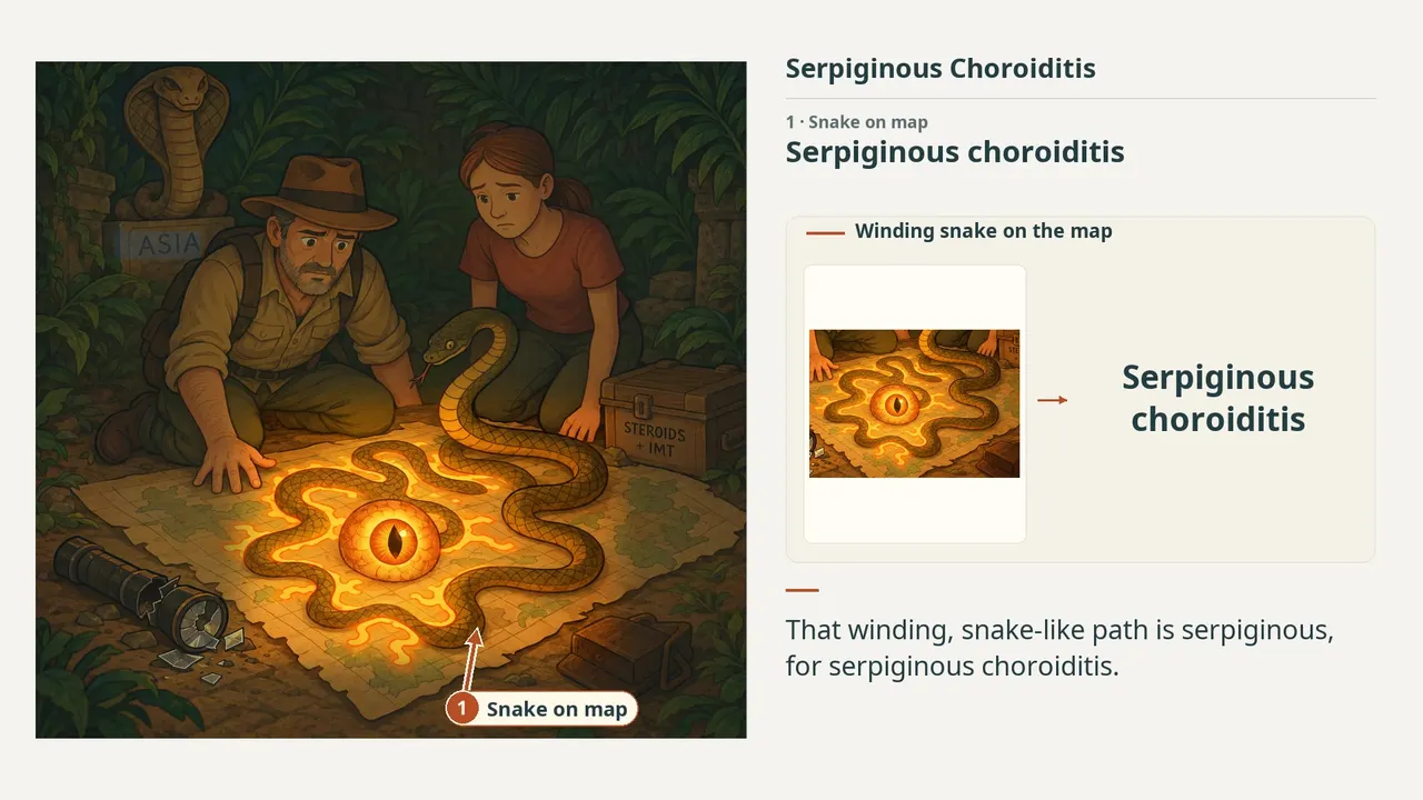
- Snake on mapSerpiginous choroiditis
- Middle-aged explorerMiddle-aged patient population
- SpreadSerpiginous, centrifugal spread of lesions
- Two explorers1:1 male:female
- Broken flashlightPoor visual-acuity prognosis
- Looping snakeRecurrent and progressive disease
- Medicine chestSystemic immunosuppression: steroids to IMT
- CobraSerpiginous-like choroiditis (TB) on the differential
- ReviewEvery marker, in order
Transcript
The narration, grouped by cue. Select a sentence to play from there.
Suggest an addition
Know a high-yield fact that deserves an anchor in this image, or spotted a mistake? Only the AfterImage team sees what you send.
Clinical image credits
Openly licensed photographs used in this lesson, with what was changed.
Mauschitz Matthias M, Zeller Markus, Sagar Pradeep, Biswal Suchitra, Guzman Gabriela, Terheyden Jan H, Meyer Carsten H, Holz Frank G, Heinz Carsten, Pleyer Uwe, Finger Robert P, Wintergerst Maximilian W M, Ferreras Antonio. Fundus Autofluorescence in Posterior and Panuveitis—An Under-Estimated Imaging Technique: A Review and Case Series. Figure 4, serpiginous crop.
Exact RGB rectangular crop [0, 0, 459, 410] from original 729 × 821 image; no enlargement, retouching, new arrows, color adjustment or generative processing. Original markings within crop preserved.