Cornea
Epithelial basement membrane dystrophy
EBMD
1:407 visual cues2 clinical photos
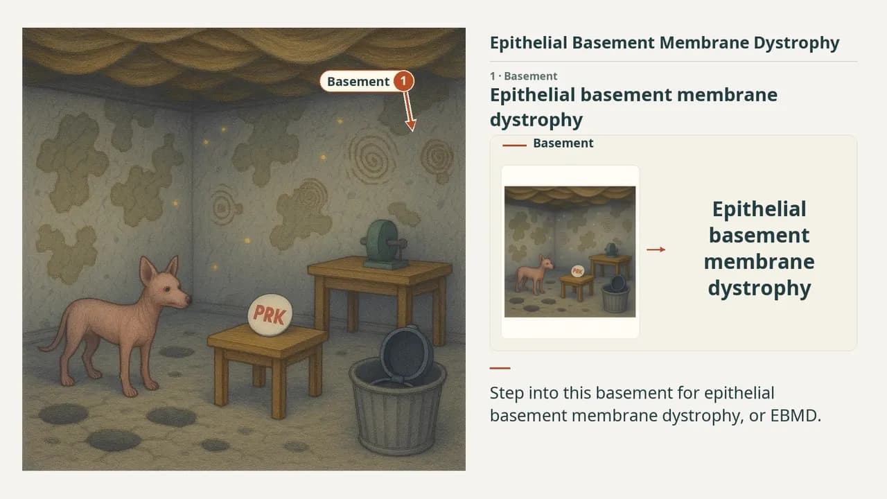
In this image
Each number marks an object in the image and the finding it stands for.
- BasementEpithelial basement membrane dystrophy
- Hairless dogNo inheritance pattern
- Floor erosionsRecurrent corneal erosions
- Redundant ceilingExtra basement membrane sheets
- Maps, dots, fingerprintsMap, dot and fingerprint findings
- PRK pinPRK: refractive surgery of choice
- LASIK kit in garbageLASIK not recommended
- ReviewEvery marker, in order
Transcript
The narration, grouped by cue. Select a sentence to play from there.
Suggest an addition
Know a high-yield fact that deserves an anchor in this image, or spotted a mistake? Only the AfterImage team sees what you send.
Clinical image credits
Openly licensed photographs used in this lesson, with what was changed.
Juliette Buffault, Pierre Zéboulon, Hong Liang, Anthony Chiche, Jade Luzu, Mathieu Robin, Ghislaine Rabut, Marc Labetoulle, Antoine Labbé, Christophe Baudouin (2020). Assessment of corneal epithelial thickness mapping in epithelial basement membrane dystrophy.
Exact rectangular crop [0, 0, 746, 287] from 746 × 907 publisher figure. Decoded RGB pixels preserved; no enhancement, arrows, retouching or upscaling.
Juliette Buffault, Pierre Zéboulon, Hong Liang, Anthony Chiche, Jade Luzu, Mathieu Robin, Ghislaine Rabut, Marc Labetoulle, Antoine Labbé, Christophe Baudouin (2020). Assessment of corneal epithelial thickness mapping in epithelial basement membrane dystrophy.
Exact rectangular crop [0, 0, 762, 342] from 762 × 342 publisher figure. Decoded RGB pixels preserved; no enhancement, arrows, retouching or upscaling.