Uveitis and white dot syndromes
Fuchs heterochromic iridocyclitis
2:5610 visual cues2 clinical photos
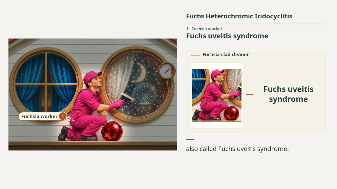
In this image
Each number marks an object in the image and the finding it stands for.
- Fuchsia workerFuchs uveitis syndrome
- Affected windowUsually unilateral
- Faded curtainHeterochromia from iris stromal atrophy
- StarsSmall stellate keratic precipitates
- Calm workerChronic, low-grade anterior uveitis
- RubyChronic rubella association
- MoonPosterior subcapsular cataract
- GaugeElevated IOP; secondary glaucoma
- Snow globeVitreous cells and opacification
- Red vinesFragile vessels; hyphema; Amsler sign
- ReviewEvery marker, in order
Transcript
The narration, grouped by cue. Select a sentence to play from there.
Suggest an addition
Know a high-yield fact that deserves an anchor in this image, or spotted a mistake? Only the AfterImage team sees what you send.
Clinical image credits
Openly licensed photographs used in this lesson, with what was changed.
Francisco J. Muñoz-Negrete, Javier Moreno-Montañés, Paula Hernández-Martínez, Gema Rebolleda (2015). Current Approach in the Diagnosis and Management of Uveitic Glaucoma. Figure 3.
Exact rectangular crop [10, 10, 726, 240] from the 736 × 278 publisher original (Figure 3), preserving decoded RGB pixels. The authors' own arrows and markings are kept; no other changes, retouching, enhancement or upscaling.
Yu Yoneda, Yoshihiko Usui, Rie Tanaka, Keitaro Hase, Kenichi Namba, Koju Kamoi, Hiroshi Takase, Masaki Takeuchi, Wataru Matsumiya, Sentaro Kusuhara, Atsunobu Takeda, Nobuyo Yawata, Ryoji Yanai, Tomona Hiyama, Yosuke Harada, Noriyasu Hashida, Kazuichi Maruyama, Kei Nakai, Ryo Taguchi, Toshikatsu Kaburaki, Nobuhisa Mizuki, Hiroshi Goto, Yujiro Fujino, Masaru Takeuchi (2022). Factors associated with low prevalence of Fuchs' uveitis syndrome in Japan. Figure 1A.
Exact rectangular crop [0, 0, 827, 619] from 827 × 948 publisher original, preserving decoded RGB pixels. No other changes, arrows, retouching, enhancement or upscaling.