Cornea
Granular corneal dystrophy type 1
1:457 visual cues2 clinical photos
In this image
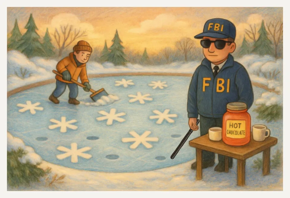
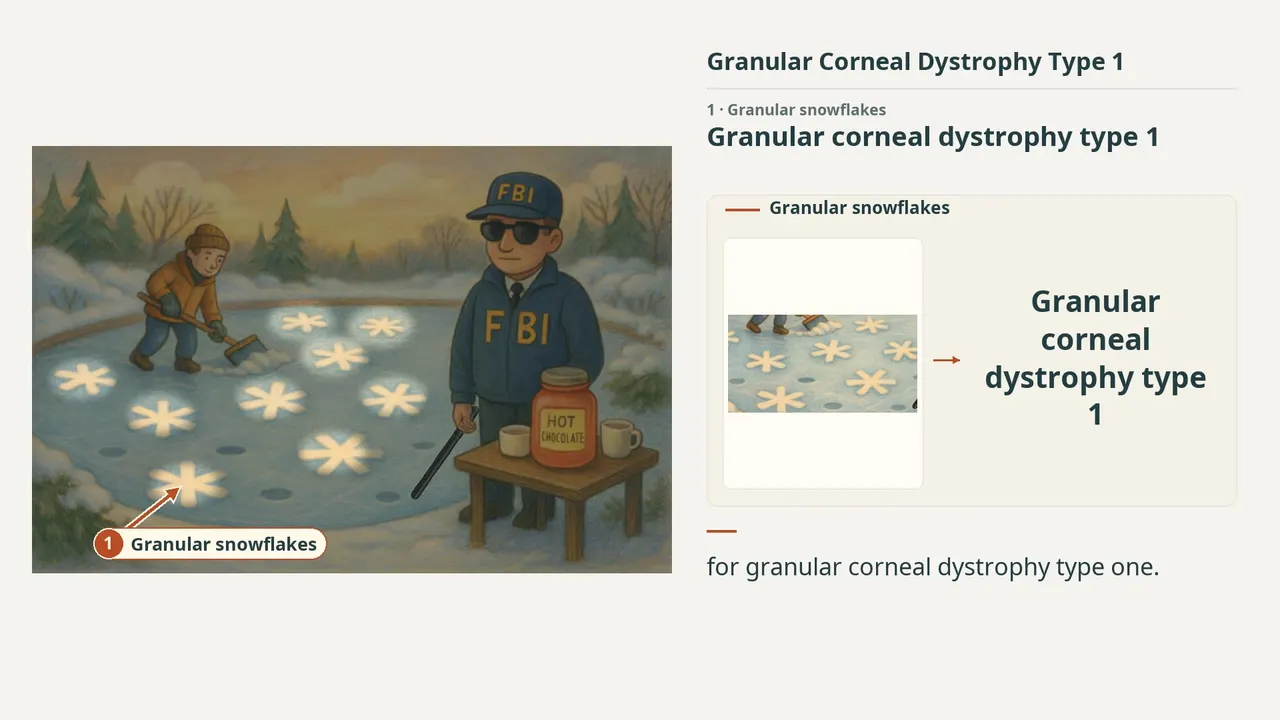
Each number marks an object in the image and the finding it stands for.
- Granular snowflakesGranular corneal dystrophy type 1
- FBI agentTGFBI gene mutation
- Clear spacesGranular deposits with clear spaces between lesions
- Clearing the edgeLesions do not extend limbus to limbus
- Red mason jarHyaline material stains red with Masson trichrome
- Ice holesRecurrent corneal erosions possible
- BatonRod-shaped fibers on electron microscopy
- ReviewEvery marker, in order
Transcript
The narration, grouped by cue. Select a sentence to play from there.
Suggest an addition
Know a high-yield fact that deserves an anchor in this image, or spotted a mistake? Only the AfterImage team sees what you send.
Clinical image credits
Openly licensed photographs used in this lesson, with what was changed.
Eduardo Gayger Müller, José Álvaro Pereira Gomes, Walton Nosé, Ricardo Menon Nosé (2018). Persistence of granular corneal dystrophy type-1 deposits in the predescemetic layer after big-bubble deep anterior lamellar keratoplasty.
Exact rectangular crop [0, 0, 900, 881] from 900 × 881 publisher figure. Decoded RGB pixels preserved; no enhancement, arrows, retouching or upscaling.
Eduardo Gayger Müller, José Álvaro Pereira Gomes, Walton Nosé, Ricardo Menon Nosé (2018). Persistence of granular corneal dystrophy type-1 deposits in the predescemetic layer after big-bubble deep anterior lamellar keratoplasty.
Exact rectangular crop [0, 0, 900, 736] from 900 × 736 publisher figure. Decoded RGB pixels preserved; no enhancement, arrows, retouching or upscaling.