Cornea
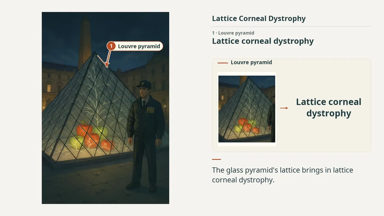
Lattice corneal dystrophy
1:366 visual cues2 clinical photosFree lesson
In this image
Each number marks an object in the image and the finding it stands for.
- Louvre pyramidLattice corneal dystrophy
- FBI agentTGFBI gene mutation
- Branching linesThin, branching, refractile central lines; Do not extend to the limbus
- Lower hazeInitially clear stroma; Haze develops over time
- Red-green applesAmyloid; Congo red-positive birefringence
- Floor cracksRecurrent corneal erosions
- ReviewEvery marker, in order
Transcript
The narration, grouped by cue. Select a sentence to play from there.
Suggest an addition
Know a high-yield fact that deserves an anchor in this image, or spotted a mistake? Only the AfterImage team sees what you send.
Clinical image credits
Openly licensed photographs used in this lesson, with what was changed.
Fengjiao Zhu, Ming Li, Chun Zhang, Chan Chen, Fangwei Ying, Danyao Nie (2021). In vivo confocal microscopy qualitative investigation of the relationships between lattice corneal dystrophy deposition and corneal nerves.
Exact rectangular crop [0, 0, 595, 460] from 595 × 460 publisher figure. Decoded RGB pixels preserved; no enhancement, arrows, retouching or upscaling.
Matsumoto, Fukuoka, Sotozono (2025). Descemet's Membrane Detachment During Cataract Surgery in Lattice Corneal Dystrophy Type I: Histopathological Analysis of Posterior Corneal Involvement. Figure 4C.
Exact rectangular crop [0, 250, 374, 530] from the 750 × 530 publisher original (Figure 4C), preserving decoded RGB pixels. The authors' own arrows and markings are kept; no other changes, retouching, enhancement or upscaling.
