Cornea
Thiel-Behnke corneal dystrophy
TBCD
1:597 visual cues3 clinical photos
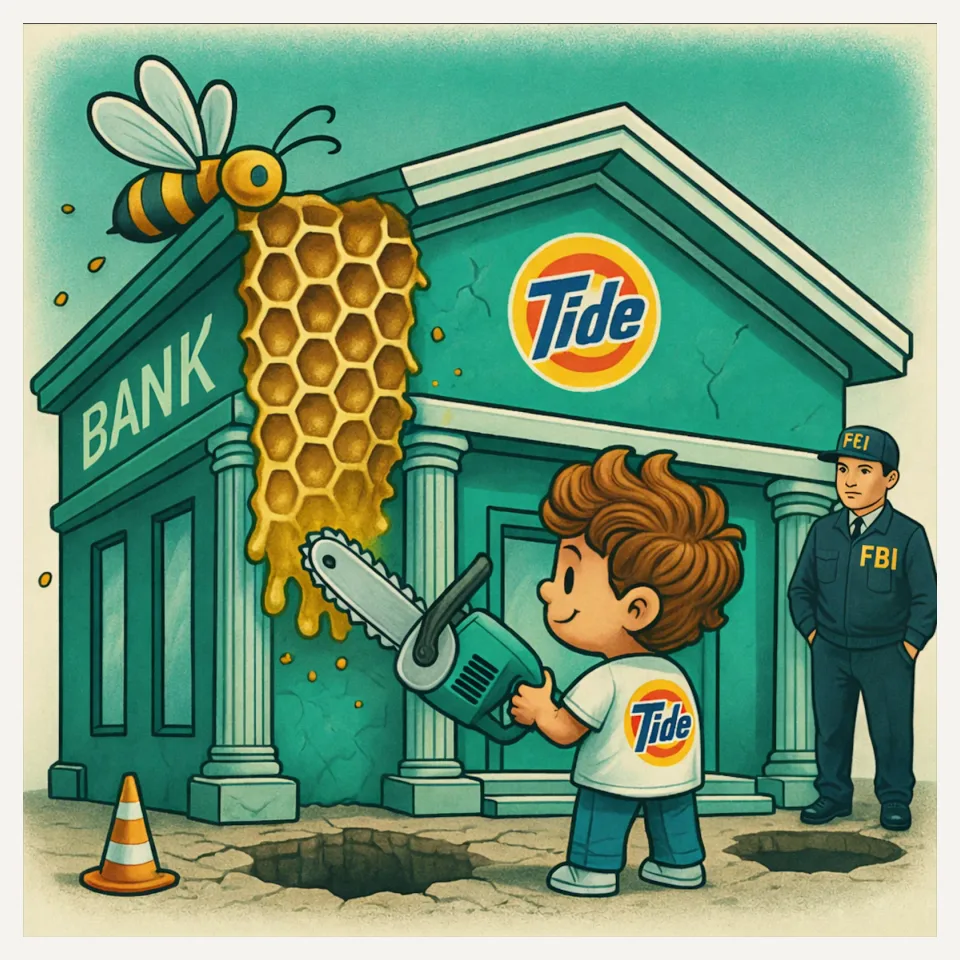
In this image
Each number marks an object in the image and the finding it stands for.
- Teal bankThiel-Behnke corneal dystrophy
- FBI agentTGFBI gene mutation
- Soft honeycombSubepithelial honeycomb opacification; Softer borders than Reis-Bücklers
- Clean Tide shirtNo amyloid / Masson trichrome staining
- ChainsawSaw-toothed Bowman’s layer; Light microscopy
- Curly hairCurly fibers; Electron microscopy
- Ground holesRecurrent epithelial erosions common
- ReviewEvery marker, in order
Transcript
The narration, grouped by cue. Select a sentence to play from there.
Suggest an addition
Know a high-yield fact that deserves an anchor in this image, or spotted a mistake? Only the AfterImage team sees what you send.
Clinical image credits
Openly licensed photographs used in this lesson, with what was changed.
Sara Bøgelund Rasmussen and Anders Peter Søndergaard (2024). Thiel-Behnke Corneal Dystrophy in a Young Man in Denmark—A Case Report.
Exact rectangular crop [0, 0, 1181, 886] from 1181 × 886 publisher figure. Decoded RGB pixels preserved; no enhancement, arrows, retouching or upscaling.
Sara Bøgelund Rasmussen and Anders Peter Søndergaard (2024). Thiel-Behnke Corneal Dystrophy in a Young Man in Denmark—A Case Report.
Exact rectangular crop [4, 5, 905, 727] from 1827 × 733 publisher figure. Decoded RGB pixels preserved; no enhancement, arrows, retouching or upscaling.
Sara Bøgelund Rasmussen and Anders Peter Søndergaard (2024). Thiel-Behnke Corneal Dystrophy in a Young Man in Denmark—A Case Report.
Exact rectangular crop [924, 4, 1816, 900] from 1827 × 909 publisher figure. Decoded RGB pixels preserved; no enhancement, arrows, retouching or upscaling.