Cornea
Lisch epithelial corneal dystrophy
LECD
1:547 visual cues1 clinical photo
In this image
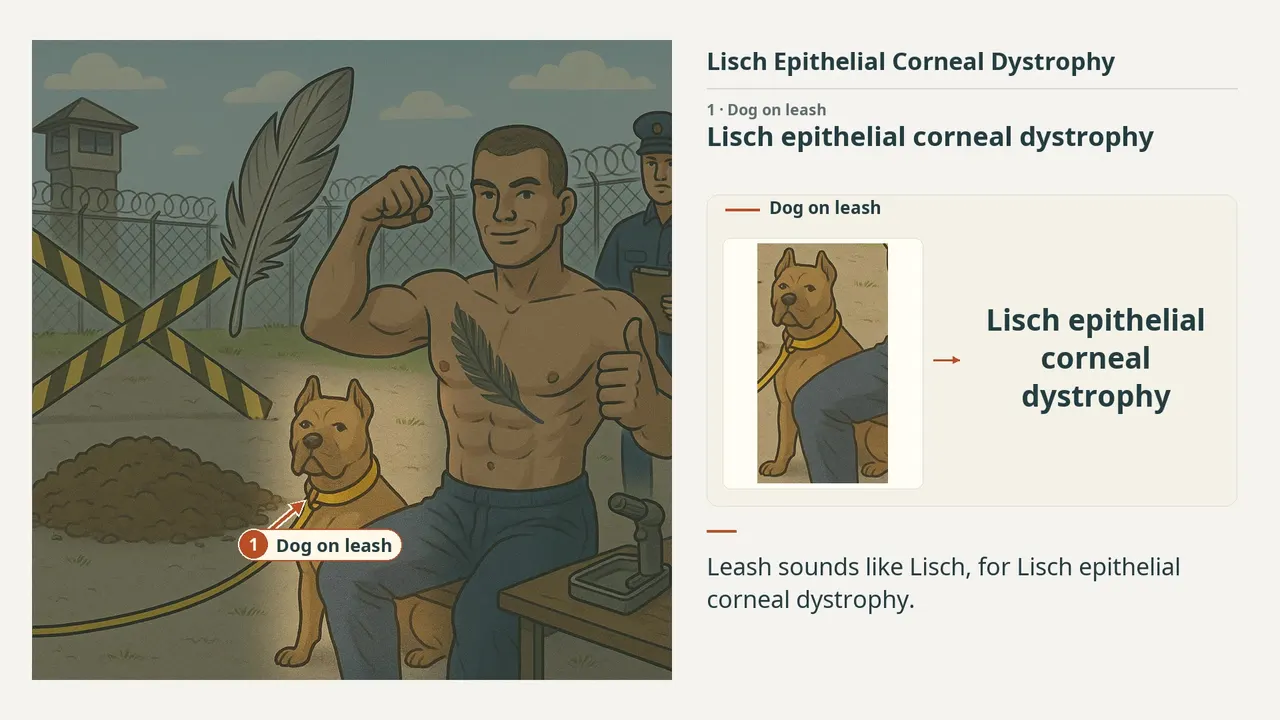
Each number marks an object in the image and the finding it stands for.
- Dog on leashLisch epithelial corneal dystrophy
- Dominant XX-linked dominant inheritance
- Flexed musclesEpithelial hyperplasia and thickening; Not microcysts
- Feather and tattooAsymmetric feathery, whorled; Gray-white epithelial opacities
- Smile and thumbs upUsually asymptomatic; Good prognosis
- Filled holeNo recurrent epithelial erosions
- Burr toolLubrication; Superficial keratectomy / PTK if visually significant
- ReviewEvery marker, in order
Transcript
The narration, grouped by cue. Select a sentence to play from there.
Suggest an addition
Know a high-yield fact that deserves an anchor in this image, or spotted a mistake? Only the AfterImage team sees what you send.
Clinical image credits
Openly licensed photographs used in this lesson, with what was changed.
Arturo E. Grau, Sergio González, Pablo Zoroquiain, Pablo A. González, Daniela Khaliliyeh, Eugenia Morselli, Dennis Cortés (2020). Evidence of autophagic vesicles in a patient with Lisch corneal dystrophy.
Exact rectangular crop [0, 0, 882, 1019] from 2700 × 2041 publisher figure. Decoded RGB pixels preserved; no enhancement, arrows, retouching or upscaling.