Cornea
Posterior amorphous corneal dystrophy
2:2812 visual cues2 clinical photos
In this image
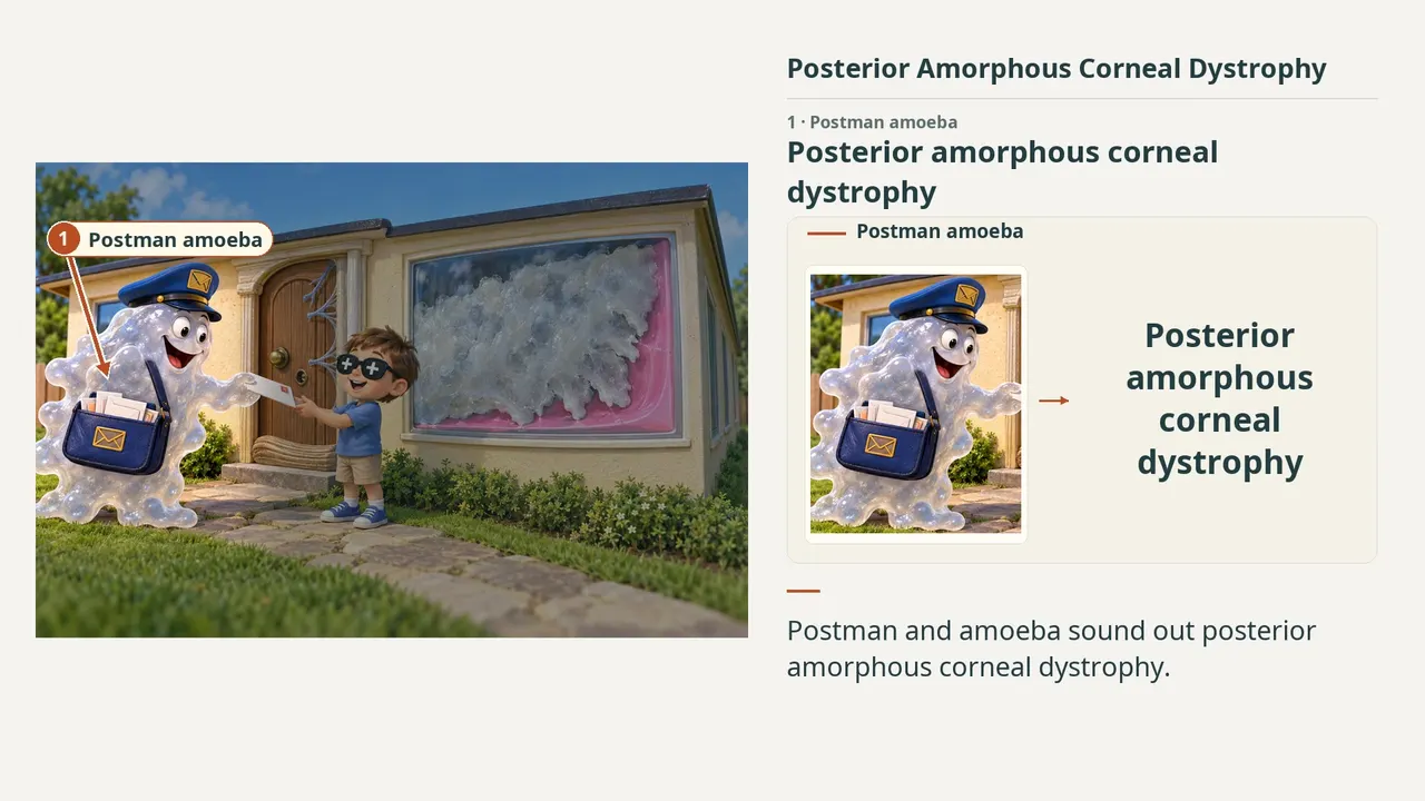
Each number marks an object in the image and the finding it stands for.
- Postman amoebaPosterior amorphous corneal dystrophy
- Posterior sheetDiffuse sheetlike posterior opacity
- Pink membraneDescemet membrane; PAS positive
- Indented layerMay indent Descemet / endothelium; Fibrillar layer can interrupt Descemet
- Young childTypically first decade; Usually nonprogressive
- Flat roofFlat cornea; Typically less than 41 D
- Thin houseThin cornea
- Plus glassesHyperopia
- Doorway ridgeProminent Schwalbe line
- StrandsFine iris processes; Iridocorneal adhesions
- Off-center knobCorectopia
- False holePseudopolycoria
- ReviewEvery marker, in order
Transcript
The narration, grouped by cue. Select a sentence to play from there.
Suggest an addition
Know a high-yield fact that deserves an anchor in this image, or spotted a mistake? Only the AfterImage team sees what you send.
Clinical image credits
Openly licensed photographs used in this lesson, with what was changed.
Michelle J. Kim, Ricardo F. Frausto, George O. D. Rosenwasser, Tina Bui, Derek J. Le, Edwin M. Stone, Anthony J. Aldave (2014). Posterior Amorphous Corneal Dystrophy Is Associated with a Deletion of Small Leucine-rich Proteoglycans on Chromosome 12.
Exact rectangular crop [1028, 0, 2066, 693] from 2066 × 1379 publisher figure. Decoded RGB pixels preserved; no enhancement, arrows, retouching or upscaling.
Michelle J. Kim, Ricardo F. Frausto, George O. D. Rosenwasser, Tina Bui, Derek J. Le, Edwin M. Stone, Anthony J. Aldave (2014). Posterior Amorphous Corneal Dystrophy Is Associated with a Deletion of Small Leucine-rich Proteoglycans on Chromosome 12.
Exact rectangular crop [1028, 693, 2066, 1379] from 2066 × 1379 publisher figure. Decoded RGB pixels preserved; no enhancement, arrows, retouching or upscaling.